



On Exhibit in the Sigel Gallery at the Falmouth Art Center from September 1-30
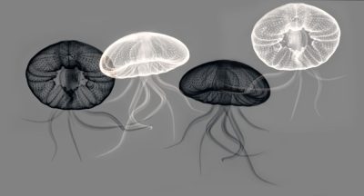
In this exhibit exhibit of X-rays as art, Dr. George Taylor of Falmouth and Philadelphia invites viewers to explore the inner world of flowers and sea creatures.
Dr. Taylor said he began to experiment with x-ray art in the mid-1990s while working as a radiologist at Johns Hopkins Hospital.
The National Aquarium, located a few blocks away in the Inner Harbor of Baltimore brought in a live chambered nautilus to see if it had enough gravel in its mantle to grind food.
Dr. Taylor said, “The CT scan showed the beautiful inner chambers only hinted at from the surface. At that moment, I knew that there were wonderful possibilities to explore as an artist using Roentgen’s invisible light,” referring to the German physicist who discovered X-rays in 1895 and won the first Nobel Prize in Physics.
At first, Dr. Taylor did straight X-ray “photograms” of flowers, shells and other small objects with little if any digital modification. These evolved into more complex collages, and eventually, into abstract visions of form and structure.
What is “x-ray art”?
Dr. Taylor explains, “X-Rays are high-energy electromagnetic waves that are invisible to the naked eye. Unlike visible light that reflects off the surface of an object, X-rays can penetrate that object, creating shadows that allows us to see its internal structure.”
He continues,” In the visible world, we usually respond to a flower because of its bright and distinct colors. As X-rays transilluminate a flower, we are able to see inside, and the flower’s framework becomes its most noticeable feature. One begins to notice how an elegance of design is repeated in both the plant and animal kingdom.”
To give an example, Dr. Taylor discusses fish and seeds. “Radial symmetry is present in the shape of a starfish. In a very similar way, it is used to maximize the number of seeds packed in a poppy seedpod. Under the X-ray, common objects take on a new identity. A flower becomes a puff of smoke, or an underwater creature swirling with its partners in an underwater ballet. By minimizing color, x-rays enhance the strong contours and forms that are otherwise not obvious to the naked eye.”
Dr. Taylor was trained as a pediatrician and a radiologist. He combined the two specialties as a pediatric radiologist, now at Children’s Hospital of Philadelphia. He is currently John A Kirkpatrick Professor Emeritus of Radiology (Pediatrics) at Harvard Medical School.
Dr. Taylor has been making photographs since 1993 and has eclectic interests, including landscapes, street and travel photography. His images have been exhibited at Lincoln Center and The Boston Museum of Science.
“As a pediatric radiologist, I spend much of my time using X-rays to detect diseases in children. Although life-saving and necessary, there is little beauty to be found in these radiographs. By using the same tools to discover hidden beauty in a flower or a shell, I can feed my soul,” he said.
Dr. Taylor will give a talk about his artwork on Tuesday, September 22, at 4pm at Falmouth Art Center. The talk is free and open to all.